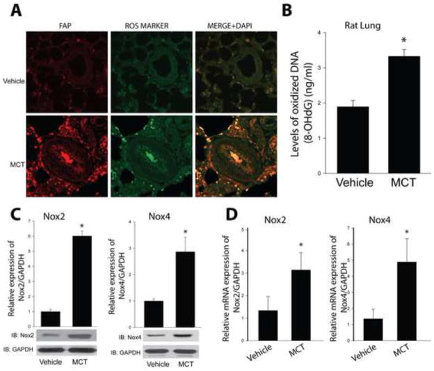
Figure 2. PH is associated with increased vascular ROS production and Nox2 and Nox4 expression.
(A) Localization of in vivo reactive oxygen species (ROS) in pulmonary arterioles. Sections of control and 4wk MCT-treated rat lungs were co-stained for fibroblast activation protein (FAP), 8 Hydroxy-2′dexoyguanosine (ROS marker) and DAPI. (B) ROS production was quantified by an Oxidative DNA Damage ELISA kit using genomic DNA isolated from control and 4wk MCT-treated rat lungs. Nox2 and Nox4 protein (C) and mRNA (D) expression were measured in isolated PA from MCT rats. Results are representative of at least 3–5 separate experiments. * different from Vehicle, p < 0.05 (n = 5–6).

