
Figure 5. Cav-1 alters the expression of Nox proteins in native cells.
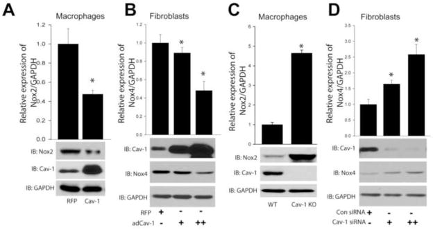
(A) Macrophages were transduced with either Cav-1 adenovirus (30MOI) or a non-specific control gene (RFP) (30MOI) for 48hrs, and cell lysates were immunoblotted for Nox2, Cav-1, and GAPDH. (B) Human lung fibroblasts were exposed to increasing amounts of Cav-1 adenovirus (10 and 30MOI) and cell lysates immunoblotted for Nox4, Cav-1, and GAPDH. (C) Macrophages from WT or Cav-1 knockout mice were isolated, and cell lysates immunoblotted for Nox2, Cav-1, and GAPDH. (D) Cav-1 expression was silenced using siRNA (10(+), 30(++) nM) in human lung fibroblasts, and cell lysates immunoblotted for Nox4, Cav-1, and GAPDH. Results are representative of at least 3–5 separate experiments.