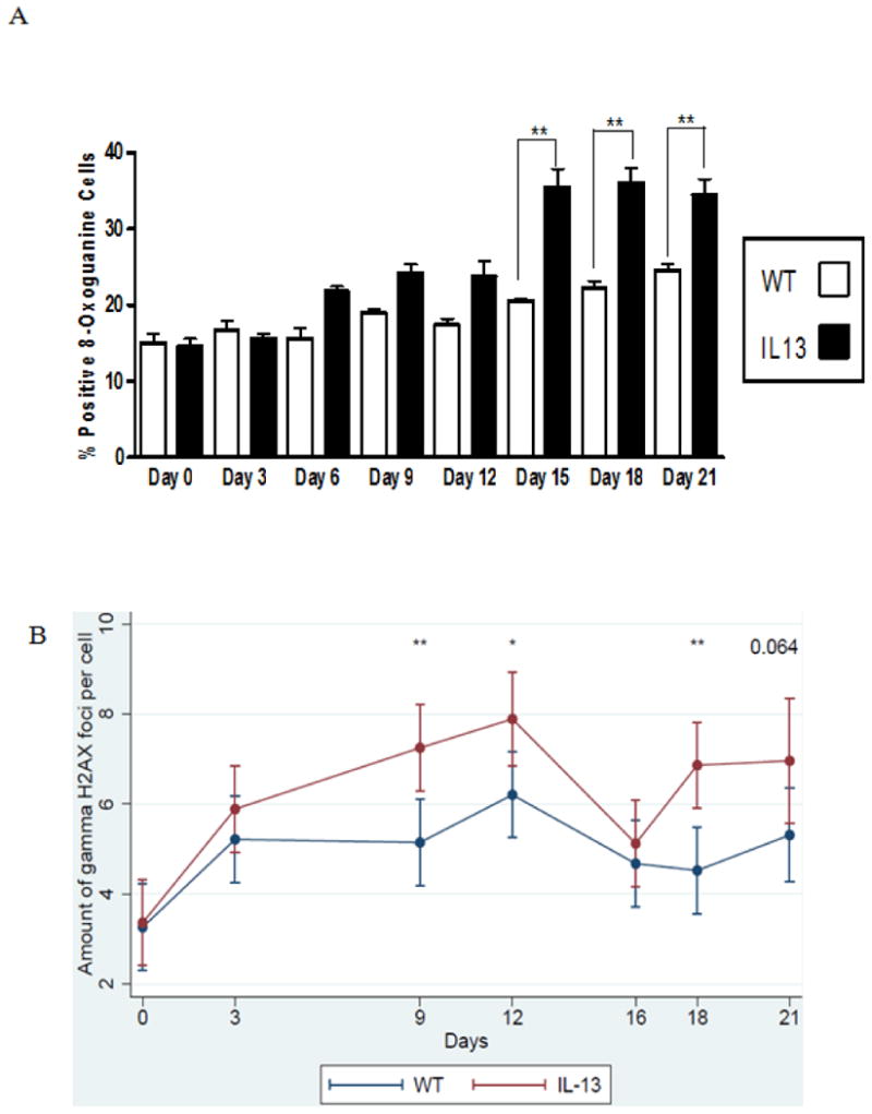
Fig.6. Persistent genotoxicity measured via inflammation induced 8-oxoguanine and double stranded breaks measured via γH2AX in peripheral blood.

A.) Percent positive cells for 8-oxoguanine induction in white blood cells. Presence of 8-oxoguanine was confirmed by immunofluorescence. Positive cells stain brightly green compared to no immunofluorescent staining for negative cells. White bars indicate Wild type (WT) animals and black bars indicate IL-13 animals. Data represent mean ± SEM. Statistical analyses were done using ANOVA testing and Tukey's post hoc analysis. n=5 in all groups. ** indicates p<0.001.B Assessment of double strand breaks measured via γH2AX assay, were counted per cell using fluorescent microscopy before doxycycline administration at Day 0 and after doxycycline administration at days 3,9,12,16,18 and day 21 using a linear mixed model to determine genotoxic accumulation over time. * indicates p<0.02, ** indicates p<0.002 n=5 for WT and IL-13 animals. (color)
