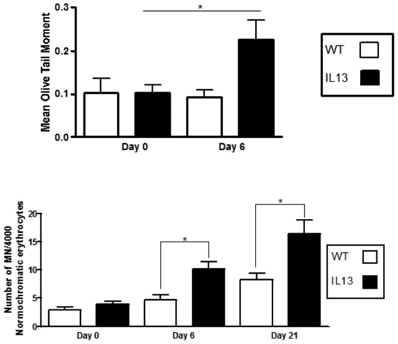
Fig.7. IL-13 over-expression induced single stranded breaks and micronucleated cells in peripheral blood.

A.) Assessment of single strand breaks were measured via comet assay before doxycycline administration at Day 0 and after doxycycline administration at days 6. At least 100 olive tail moments were counted via fluorescent microscopy and assessed using CASP software. White bars indicate Wild type (WT) animals and black bars indicate IL-13 animals. Data represent mean ± SEM. Statistical analyses were done using ANOVA testing and Tukey's post hoc analysis. * indicates p<0.05 n=5 for WT and IL-13 animals. B.) Number of micronucleated cells per 4000 normorchromatic erythrocytes. Presence of micronuclei were confirmed by light microscope at 100X. White bars indicate Wild type (WT) animals and black bars indicate IL-13 animals. Data represent mean ± SEM. Statistical analyses were done using ANOVA testing and Tukey's post hoc analysis. n=9 for WT and n=10 for IL-13.* indicates p<0.05.
