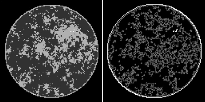
Fig. 1.

(Left) Discrete phantom modeled after a breast CT application shown in the gray-scale window  . (Right) Gradient magnitude image (GMI) of the phantom shown in the gray scale window
  . The units of the GMI are also
  , because the numerical implementation of
  involves only the differences between neighboring pixels without dividing by the physical pixel dimension. The phantom array is composed of 12,892 pixel values, and there are 4,053 non-zero values in the GMI.
