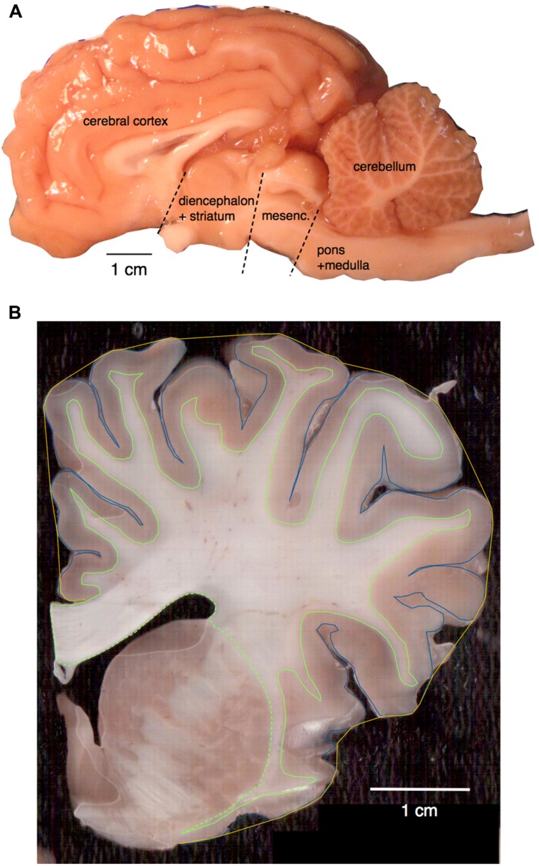
FIGURE 1.
(A) Main structures analyzed. Shown is the hemisphere of Damaliscus dorcas, illustrating the separation of pons + medulla from the mesencephalon, and the cut that separated the latter from the diencephalon. The ensemble of diencephalon + striatum were later separated from the cerebrum in the individual coronal sections cut from the hemisphere already stripped of the mesencephalon. (B) Surface areas and volumes shown on a coronal section of Giraffa camelopardalis. Total pial surface perimeter (PG) is shown in blue; exposed perimeter, in yellow; perimeter of the white–gray matter interface (PW) in green (solid line); and area of subcortical white matter in the coronal plane also in green, including the dashed line.

