Figure 1.
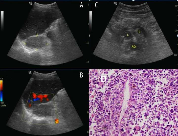
Typical ultrasound imaging of adrenal carcinoma. (A) Right adrenal mass with 6.1×3.6 cm size, middle uniform echo, and unclear boundary. (B) No notable vascularization is shown within the mass on color Doppler sonography. (C) Multiple enlarged lymph nodes were found retroperitoneally (D) Histopathology result showed adrenal adenomas.

