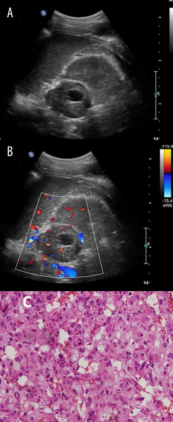
Figure 4.

Typical ultrasound imaging of pheochromocytomas with necrosis in a 31-year-old man with a 5-year history of hypertension. (A) Left adrenal mass with 5.5×4.1 cm size, middle and uneven echo, and visible liquefied area. (B) Blood flow signals were found mainly in the lesion but also around the lesion by color Doppler examination. (C) Histopathology result showed pheochromocytomas.
