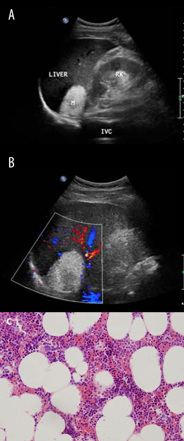
Figure 5.

Typical ultrasound imaging of myelolipoma in a 59-year-old man. (A) Right adrenal mass 52×39 mm in size and homogenous hyperechoic. (B) No notable vascularization is shown within the mass (arrows) on color Doppler sonography. (C) Histopathology result showed myelolipoma.
