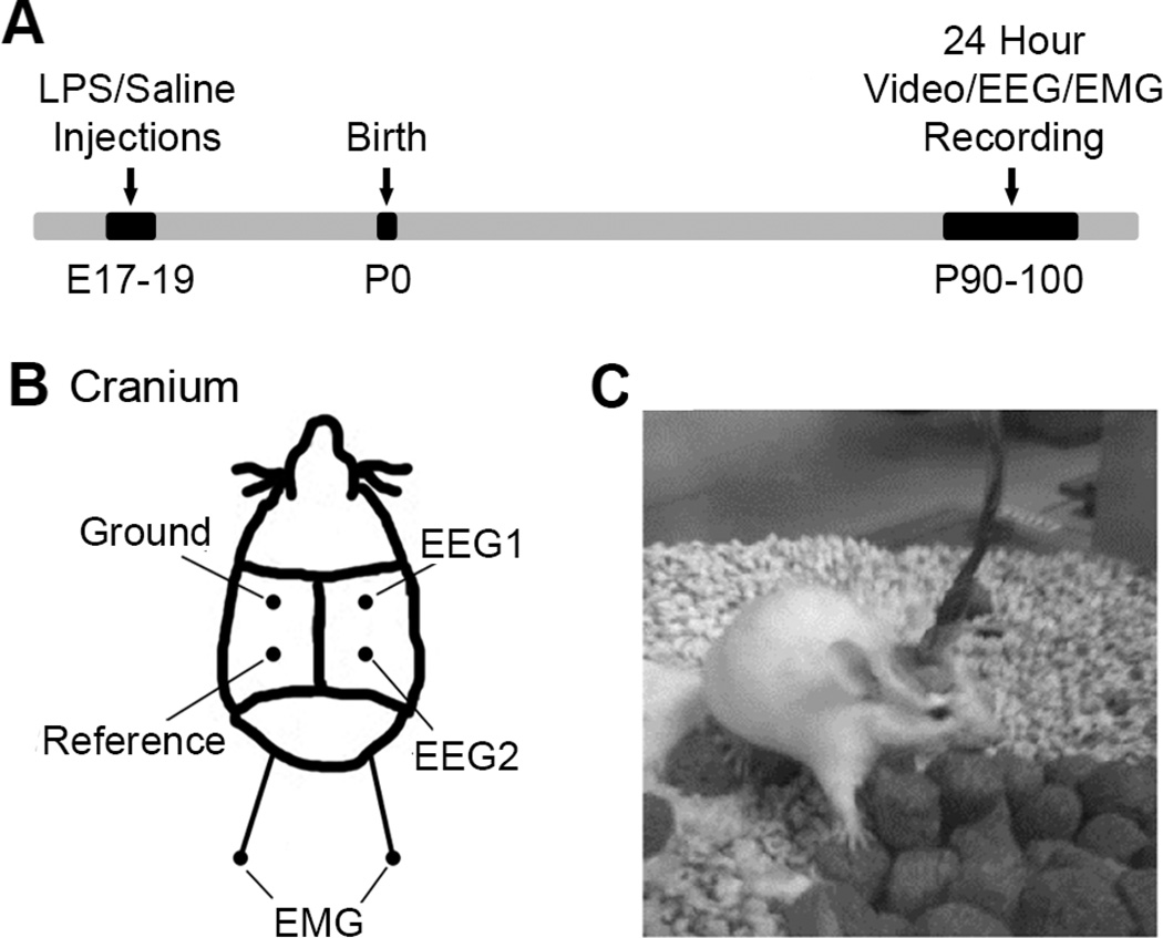
Figure 1. Schematic with experimental timeline and EEG placement.
(A) The timeline of events starting with LPS/Saline injections at prenatal 17–19, to birth and recordings at post-natal 90–100. (B) The location of the EEG/EMG electrodes on each mouse’s cranium. EEG1, EEG2, ground and reference subdural electrodes were placed parasagittally. In addition, two EMG electrodes were placed sub-dermally over supra scapular region. (C) A representative mouse during a tethered recording in recording chamber.

