
Extended Data Figure 1. The proximal remnants of truncated skeletal elements in D sagitta are correctly patterned.
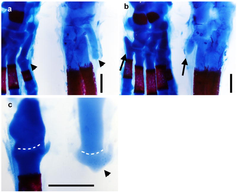
Alcian blue and alizarin red stained skeletons of postnatal day 0 mouse (left) and three-toed jerboa, D sagitta (right) with proximal (ankle) at the top. a, Anterior view highlights the first metatarsal (arrow head). b, Posterior view highlights the fifth metatarsal (arrow). c, Dissected first tarsal-metatarsal elements demonstrate the morphology of the truncated first metatarsal of D sagitta (right, arrow head) compared to mouse (left). Joint interzone indicated by white dashed line. Scalebars = 0.5 mm.