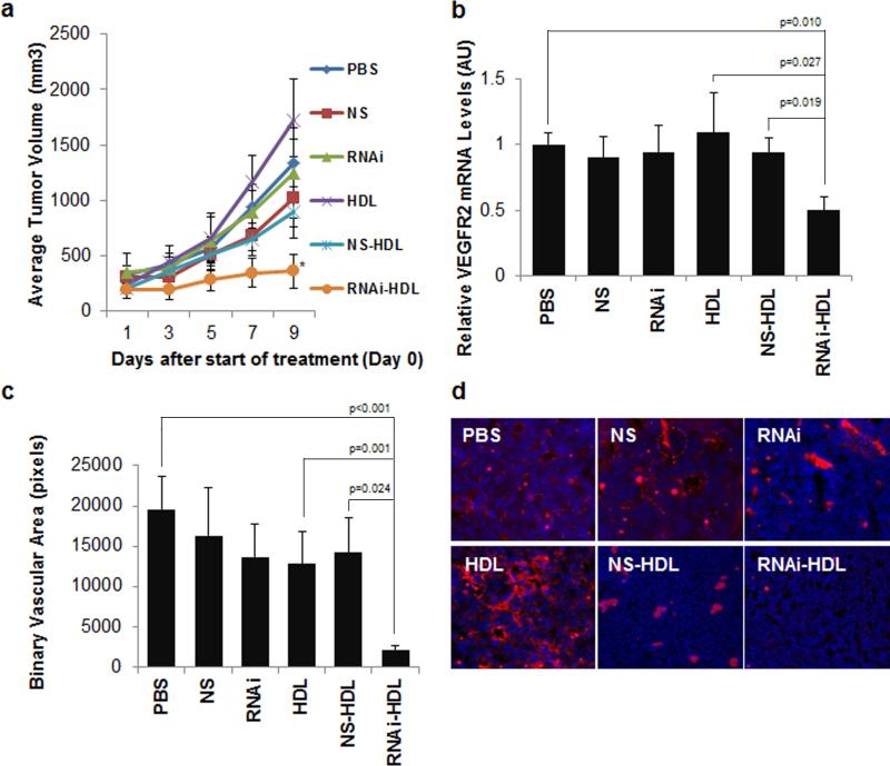
Figure 6.
Lewis lung carcinoma (LLC-1) subcutaneous tumor response to intravenous administration of RNAi-HDL NPs and control treatments. (a) Average tumor volumes are plotted for each of the treatments and there is a significant reduction in the RNAi-HDL NP (RNAi-HDL) treatment group on day 9 as compared to NS-HDL (p=0.016). (b) Tumors from the RNAi-HDL NP (RNAi-HDL) treatment group exhibit a significant reduction in VEGFR2 mRNA content relative to ß-actin mRNA. (c) Binary vascular area quantified by CD31 immunostaining and subsequent fluorescent imaging of frozen tumor sections reveals a significant reduction of neovascularization in RNAi-HDL NP treatment group as compared to controls. (d) Representative fluorescent images of tumor sections showing CD31 immunostained regions (red) and DAPI stained cell nuclei (blue).

