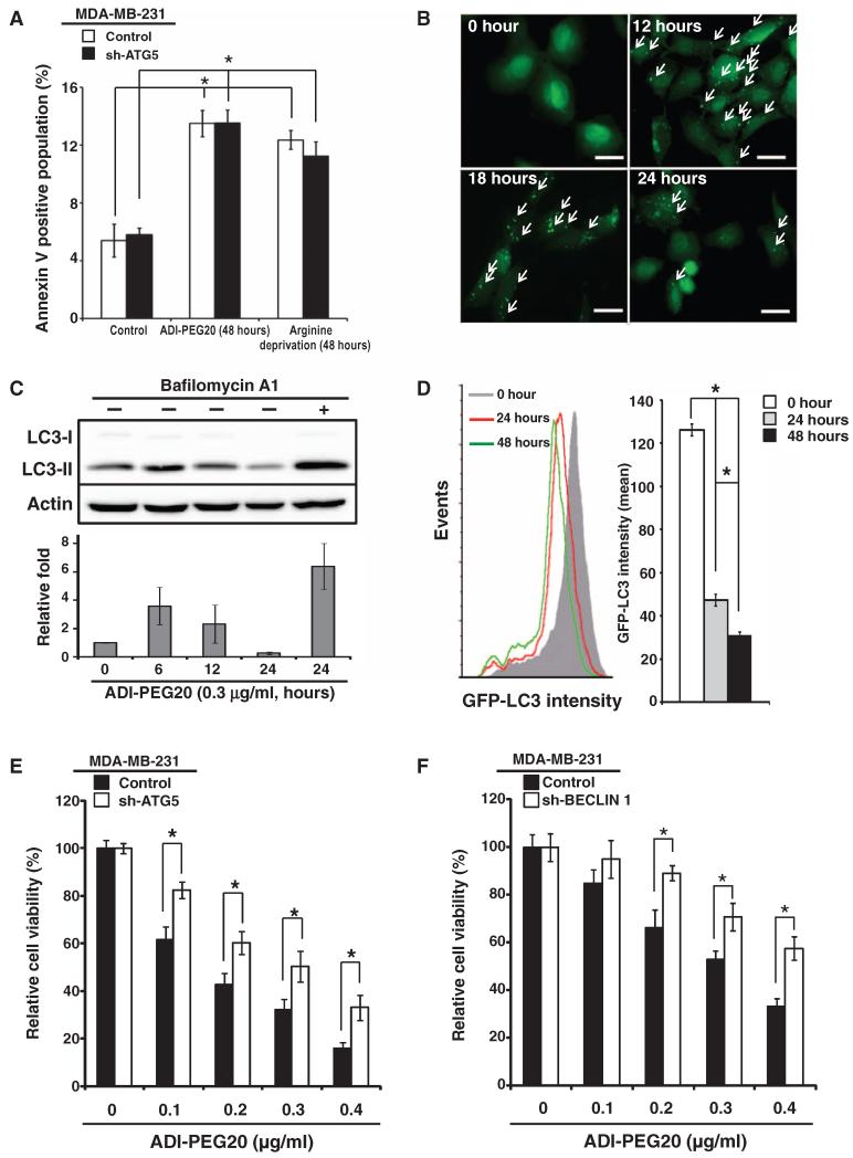
Fig. 2. ADI-PEG20 induces autophagy-dependent cell death.
(A) ADI-PEG20 treatment or arginine starvation moderately increases apoptotic cell percentage in MDA-MB-231 cells. Knockdown of ATG5 in MDA-MB-231 cells does not significantly affect response to ADI-PEG20 treatment or arginine starvation. Histograms are presented as means ± SD; n = 3 sets of cells. (B) Representative images of GFP-LC3 puncta formation (indicated by arrows) using fluorescence microscopy; n = 5 sets of cells. Scale bars, 50 mm. (C) ADI-PEG20 induces LC3 lipidation, which is reversed by bafilomycin A1. One representative Western and bar graph, presented as means ± SD, are shown; n = 3 sets of cells. (D) ADI-PEG20 induces autophagic flux. MDA-MB-231 cells overexpressing GFP-LC3 were treated with ADI-PEG20 for the indicated time periods and analyzed by flow cytometry (left panel). Data are shown as means ± SD (right panel); n = 3 sets of cells. *P < 0.05. (E and F) Knockdown of ATG5 (E) or BECLIN 1 (F) reduces ADI-PEG20-induced cytotoxicity. n = 3 sets of cells; *P < 0.05.

