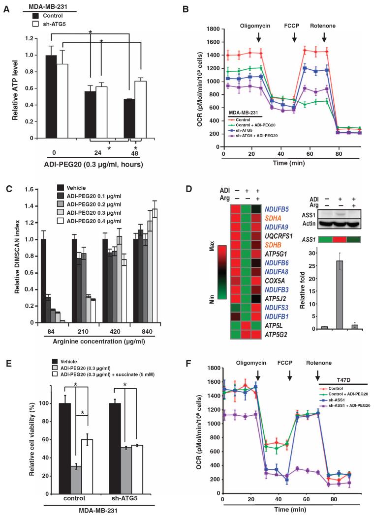
Fig. 3. ADI-PEG20 impairs mitochondrial function.
(A) Autophagy-dependent ATP concentration decreases over time in ADI-PEG20–treated MDA-MB-231 cells; n = 3 sets of cells; *P < 0.05. (B) Combination of ADI-PEG20 treatment and ATG5 gene context regulates the basal and maximal mitochondrial respiration. ATG5 knockdown partially rescued the maximal respiration stimulated by FCCP in MDA-MB-231 cells treated with ADI-PEG20; n = 3 sets of cells. OCR, oxygen consumption rate. (C) Extracellular arginine reverses the reduced cell proliferation induced by ADI-PEG20 in a dose-dependent manner; n = 5 sets of cells; *P < 0.05. (D) ADI-PEG20 treatment reversibly decreases the abundance of mRNAs encoding mitochondrial respiratory chain proteins (left panel). A list of top candidates includes messages encoding proteins of complex I (blue) and complex II (orange). Other messages shown in black are UQCRFS1 (complex III), COX5A (complex IV), and ATP5G1 and ATP5J2 (complex V). Arginine supplementation rescued ASS1 message and protein abundance in ADI-PEG20–treated MDA-MB- 231 cells (right panel), thus verifying the effectiveness of arginine supplementation. Bar graph, presented as means ± SD, from three independent Western blots is shown. ATP5L (complex V) message abundance increased in response to ADI-PEG20, and then decreased after the addition of arginine. (E) Succinate rescues the cytotoxic effect of ADI-PEG20. Data are shown as means ± SD; n = 3 sets of cells; *P < 0.05. (F) Knockdown of ASS1 suppresses basal and uncoupled mitochondrial OCR in T47D cells; n = 3 sets of cells.

