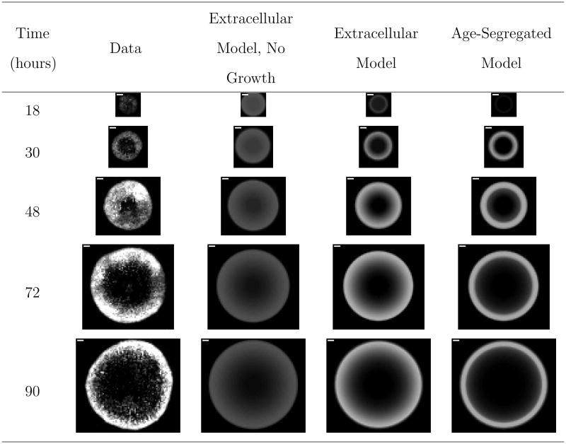
Figure 4.
Comparison of representative experimental images to model fits for VSV propagation on BHK cells. The full set of experimental images are available in Figure S1 of the supporting information. “Extracellular Model, No Growth” refers to the derived reaction-diffusion model in which the infected cells are not segregated by age and uninfected cells are not permitted to grow. “Extracellular Model.” is the same as the “Extracellular Model, No Growth” with the addition that uninfected cells are assumed to grow over the course of the experiment. “Age-Segregated Model” segregates the infected cell population by the age of infection. The white scale bar in the upper left-hand corner of the experimental images is one millimeter.

