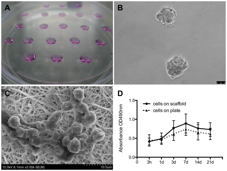
Figure 2. EB formation and the iPSC culture on the scaffold in vitro.
(A) Formation of EBs via serial dripping. Each drop of culture medium contained ∼2−5×10³ cells, which congregated and developed into an EB (B). (C) A cluster of iPSCs on the scaffold. (D) Proliferation of the iPSCs cultured on the scaffolds and on the plate was determined using CCK-8 tests; results revealed that the scaffolds promoted cell growth.

