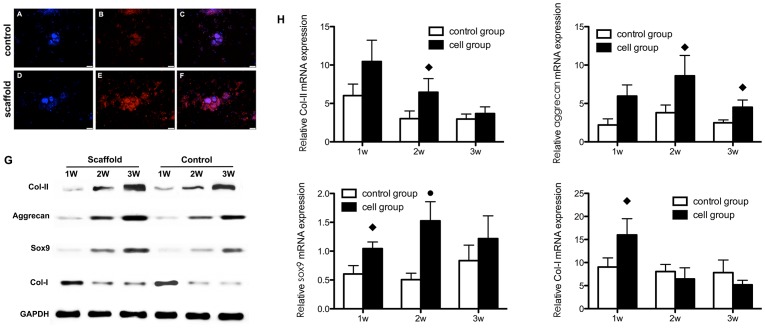
Figure 3. Chondrogenesis of iPSCs cultured on scaffolds (scaffold group) or plates (control group) in vitro.
Collagen II synthesis in the scaffold group (A,B,C) and control group (D,E,F) was analyzed by immunofluorescence staining, including nuclear counterstaining with DAPI (A,D), anti-collagen II staining in red (B,E); merged images are also shown (C,F). (G) qRT-PCR analysis of gene expression and (H) western blotting for collagen II, aggrecan, sox9 and collagen I, using GAPDH as the internal reference. The nanofibrous scaffolds generally enhanced cartilage-specific gene expression and protein levels. ♦ Statistically significant difference compared with the control group (p<0.05); • Statistically significant difference compared with the control group (p<0.01).

