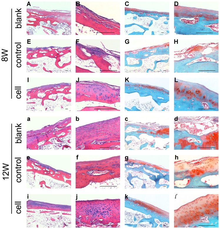
Figure 4. Histological evaluation of scaffolds and iPSCs in the restoration of cartilage defects at 8 weeks (A–L) and 12 weeks (a–l).
Blank group (A–D, a–d) defects were filled with fibrocartilage, while abundant cells were observed in the cell group (I–L, i–l). The H&E and safranin-O stained images in the right column are magnifications of images in the left column. All scale bars represent 200 µm.

