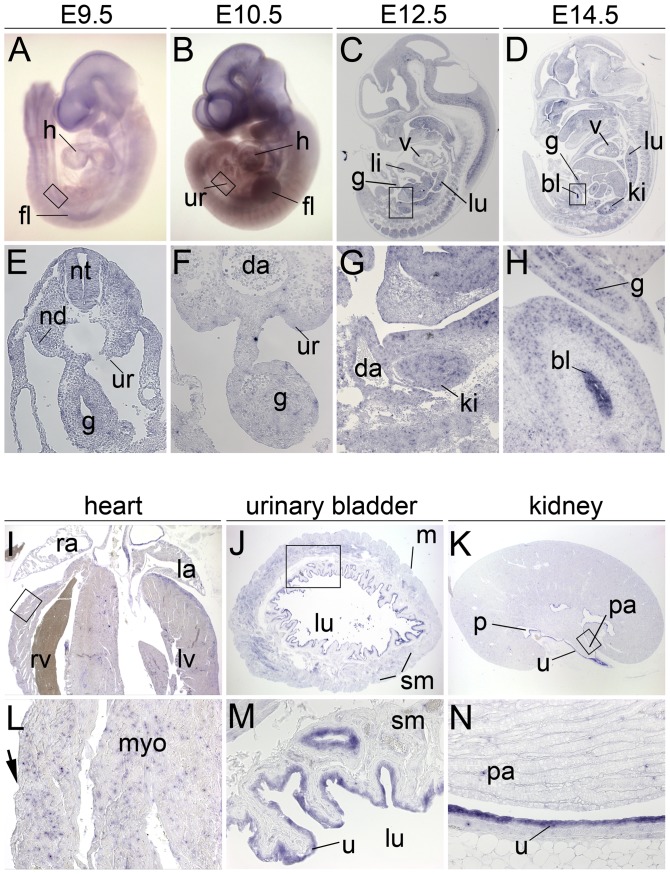
Figure 9. Upk3a expression in embryonic development and in adult tissues.
In situ hybridization analysis of Upk3a expression in whole wildtype embryos (A, B), on sagittal embryo sections (C, D and G, H), on transverse embryo sections (E, F) and on sections of the adult heart (I, L), the urinary bladder (J, M) and the kidney (K, N). (A–D) Overview images of embryos; anterior is up, dorsal is to the right. (I–K) Overview images of whole organ sections; (E–H and L–N) higher magnification images of the regions marked by open rectangles (in A–D and I–K). Stages are as indicated. Arrows point to the epicardium. bl, urinary bladder; da, dorsal aorta; fl, fore limb bud; g, gut; h, heart; ki, kidney; la, left atrium; li, liver; lu, lung; lv, left ventricle; nd, nephric duct; nt, neural tube; p, renal pelvis; pa, renal papilla; ra, right atrium; rv, right ventricle; sm, smooth muscle layer; u, urothelium; ur, urogenital ridge; v, ventricle.

