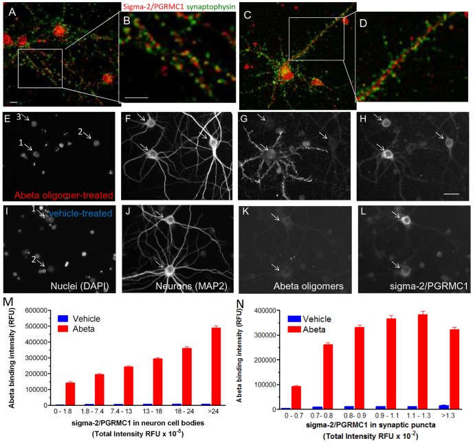
Figure 2. Sigma-2/PGRMC1 protein localizes to synaptic puncta on mature primary hippocampal cultures (21 days in vitro) and expression levels are positively correlated with Abeta oligomer binding.
sigma-2/PGRMC1 (A–D, red) is expressed at low levels in untreated cultures and is localized in cell bodies of neurons and glia, in neurite shafts, and adjacent to presynaptic puncta (A–D, synaptophysin = green) B. 66.7%±2.4 (average ± S.E.M., N = 110 neurons) of PGRMC1 positive puncta on neurons co-localize (yellow) with synaptophysin positive puncta. E–L. Positive correlation between sigma-2/PGRMC1 expression and Abeta oligomer binding in neurons (Abeta oligomers = 400 nM, 1 hour treatment). E–H. Only one neuron (MAP2 positive arrow #1 in E–H) in this field is labeled with punctate Abeta oligomer binding (G), and exhibits elevated PGRMC1 expression (H, 3.3×10⁵ RFU) compared to surrounding neurons (#2 = 1.6×10⁵, #3 = 1.8×10⁵ RFU). I–L. Vehicle-treated cultures express a similar range of sigma-2/PGRMC1 expression in neurons (arrow #1 in I = 2.62×10⁵, #2 = 1.21×10⁵ RFU). All scale bars = 20 microns. M, Binning neurons according to their intensity of sigma-2/PGRMC1 immunofluorescence and graphing the average values for Abeta binding from each bin reveals a positive correlation between the intensity of Abeta oligomer binding to synaptic puncta and the expression of sigma-2/PGRMC1 in the cell body that is significant (Kruskal-Wallis, p<0.001). N. A similar analysis of sigma-2/PGRMC1 imunofluorescence in the synaptic puncta also shows a positive correlation with Abeta oligomer binding intensity to synaptic puncta (Kruskal-Wallis p<0.001).

