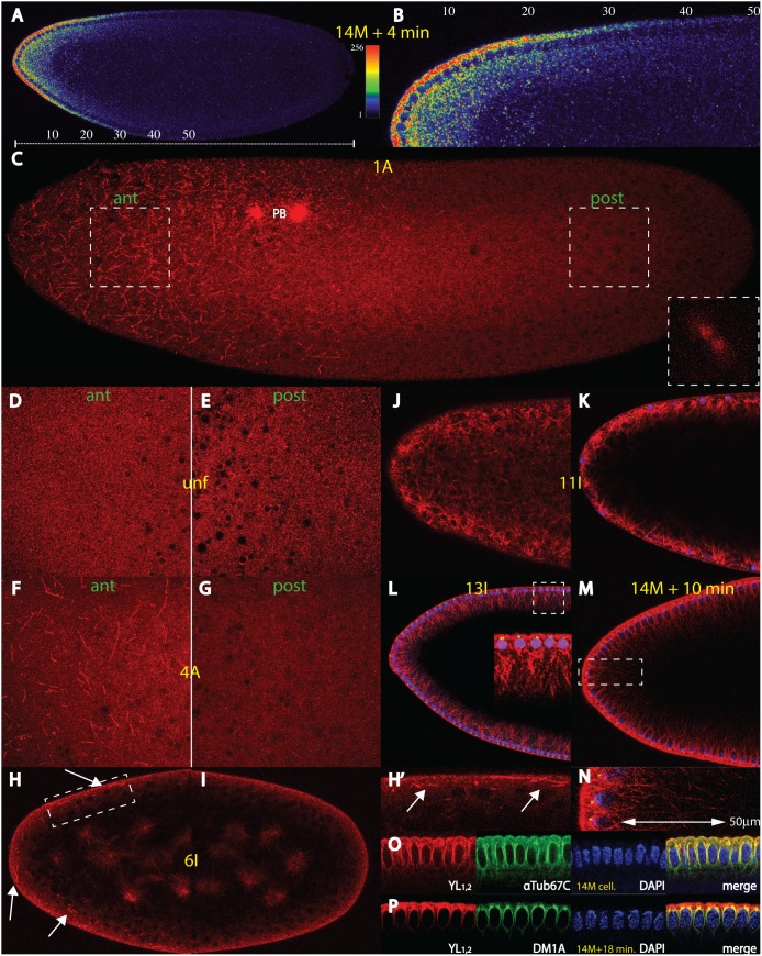
Figure 1. The bicoid mRNA gradient and an anterior cortical microtubular network.
(A) A single confocal section of a nuclear cycle (nc) 14+4 min. Drosophila embryo showing a typical bcd mRNA gradient extending up to 50% of egg length (EL, scale bar below embryo). Fluorescence intensities, reflecting bcd mRNA concentrations were converted to a colour scale shown at the right (cf. Material and Methods). (B) High magnification of the dorsal region of embryo in (A), numbers of EL above the embryo. (C) Confocal analysis at the surface of a nc 1 embryo at anaphase (insert on lower right) using mab YL1,2 against tyrosinated microtubules (MT), showing a network exclusively in the anterior half of the embryo, along with the polar bodies (PB). Two adjacent confocal sections 2.48 µm apart and maximal intensity projection were used. White areas denote magnifications of corresponding anterior (ant) regions and posterior (post) regions used in (D–G). (D) and (E) magnification of anterior (D) and posterior (E) portions on the surface of an unfertilized embryo. No network is visible. (F) and (G) magnification of anterior (F) and posterior (G) portions on the surface of an embryo at nc 4. The network is visible at the anterior half, but is absent in the posterior half. (H), (H’) and (I) sagittal confocal sections of the anterior (H) and the posterior (I) half of an embryo at nc 6. (H’) is a magnification of the area indicated in (H). The MT threads are exclusively at the anterior cortex (arrows). In the interior, asters of interphase nuclei are seen. (J) confocal section just below the nuclear layer of a nc 11 embryo and (K) mid-sagittal section of the same embryo, supplemented with the signal from the nuclear stain using DAPI (blue) and an antibody against γTubulin (green). A dense network is detected around the nuclei. (L) confocal mid-sagittal section of a nc 13 embryo, along with the signals from the nuclear stain using DAPI (blue) and an antibody against γTubulin (green). An extensive MT network ranging deeply into the yolk is visible (insert, thickness about 30 µm). (M) and (N) confocal mid-sagittal section of a nc 14+10 min. embryo, along with the signals from the nuclear stain DAPI (blue) and an antibody against Minispindles (green). (N) high magnification of the insert in (M) showing an extensive MT network ranging deeply into the yolk (insert, about 50 µm deep). (O) colocalization of mab YL1,2 to polyclonal αTub67C antibody in a cellularised nc 14 embryo, along with DAPI staining. Most YL1,2-positive threads are also positive for αTub67C. (P) colocalization of mabYL1,2 to mab DM1A in a nc 14+18 min. embryo, along with DAPI staining. Most YL1,2 threads are co-localized with DM1A at perinuclear MTs, while the periplasm is virtually free from αTub84B & D. Stages of embryos are denoted in yellow and follow the nomenclature of [61] and [3].

