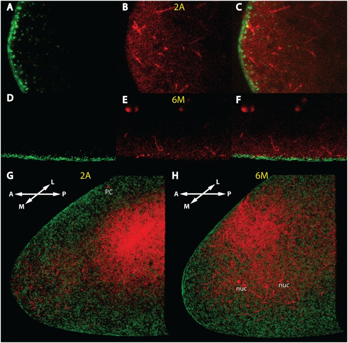
Figure 2. Independence of the early MT network from the actin sheet.
(A)–(C) mid-sagittal confocal sections of the anterior tip of a nc 2 embryo stained with Phalloidin (A) to reveal the actin structure, with mab YL1,2 against tyrosinated Tubulin (B) and merge in (C). (D)–(F) mid-sagittal section of a ventral region about 50 µm away from the anterior tip of a nc 6 embryo, stained with Phalloidin (D), mab YL1,2 (E) and merge in (F). (G) 3-D reconstruction of the confocal stack of the embryo of (A)–(C), view is from the middle (M) to the more lateral (L) part of the embryo. For film of 3D view, see Video S2. (H) 3-D reconstruction of the confocal stack of the embryo of (D)–(F), view is identical as in G. For film of 3D view, see Video S3. The red background on the inner “roof” of the embryos in (G) and (H) is excess of free tubulin which could not be removed during background subtraction of the 3D-program. Stages of embryos are denoted in yellow and follow the nomenclature of [61].

