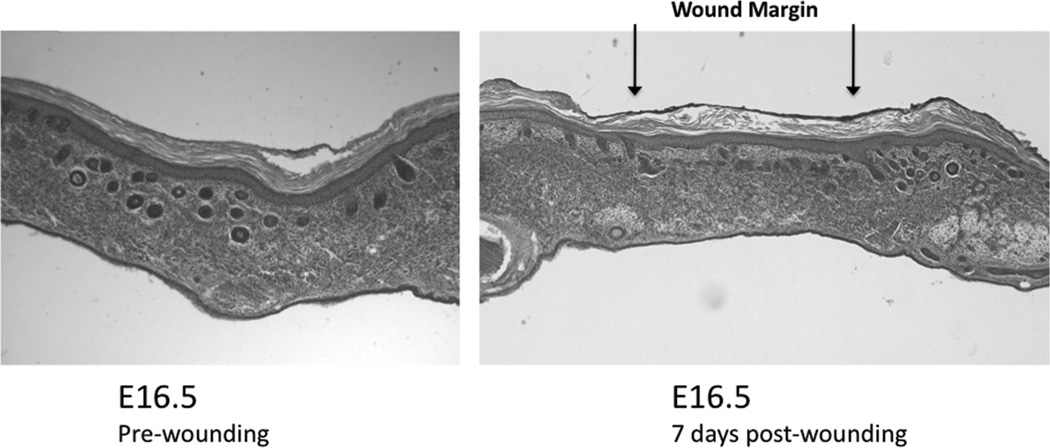
FIGURE 4.
Histology of wounded fetal skin xenografts. Normal E16.5 fetal skin (left) compared with E16.5 fetal skin transplanted onto the CAM and injured with a laser at 7 days post injury (right). The fetal skin graft wound was marked with green dye at the time of harvest. The skin wound healed with complete reepithelialization. The dermis healed with a normal extracellular matrix pattern but with a lack of rete pegs and follicular appendages. The graft skin shows little differentiation since transplantation.

