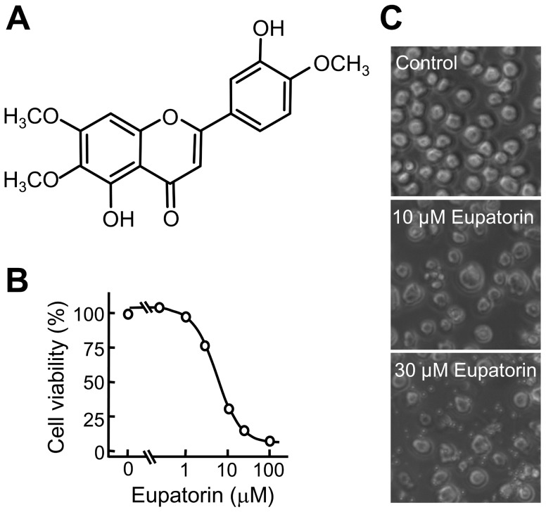
Figure 1. Chemical structure of eupatorin and its effect on human HL-60 cell viability.
(A) Structure of eupatorin. (B) Changes in cell viability as determined by the MTT assay. HL-60 cells were cultured in the presence of the indicated concentrations for 72 h, and the results are representative of those obtained in at least three independent experiments. (C) Cells were incubated with vehicle (control) or the indicated concentrations of eupatorin for 24 h and images were obtained with an inverted phase-contrast microscope.

