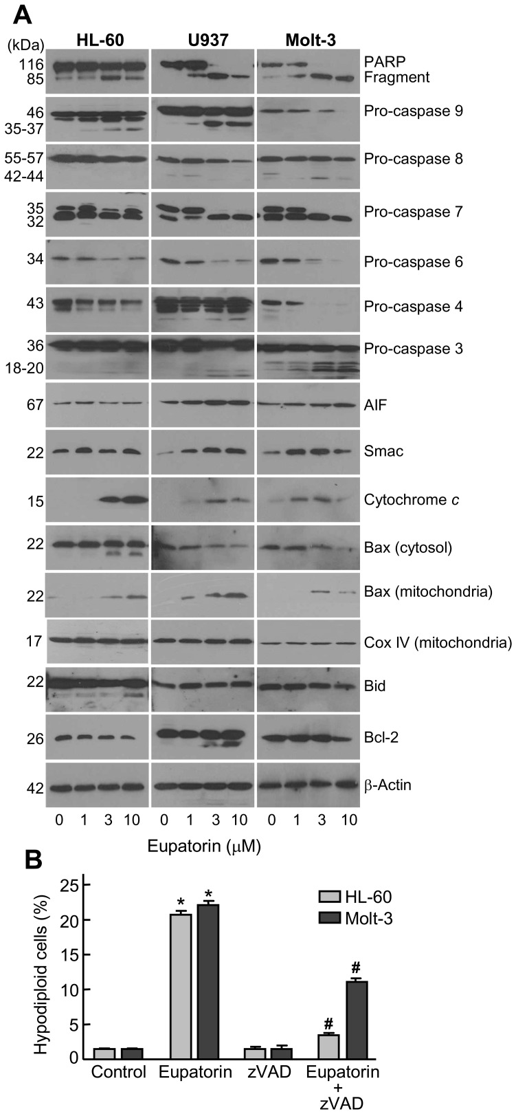
Figure 3. Involvement of caspases in the apoptosis induced by eupatorin on human leukemia cells.
(A) The cells were incubated in the presence of the indicated concentrations of eupatorin and cell lysates (or cytosolic extracts in the case of cytochrome c, AIF and Smac) were assayed by immunoblotting. β-Actin and Cox IV (cytochrome c oxidase) were used as loading controls in cytosol and mitochondria, respectively. (B) Effect of cell-permeable pan-caspase inhibitor z-VAD-fmk on eupatorin-stimulated apoptosis. HL-60 and Molt-3 cells were incubated with 10 µM eupatorin for 24 h, in absence or presence of z-VAD-fmk (100 µM) and apoptotic cells (i.e. hypodiploid DNA content) were quantified by flow cytometry after staining with propidium iodide. * indicates P<0.05 for comparison with untreated control, # indicates P<0.05 for comparison with eupatorin treatment alone.

