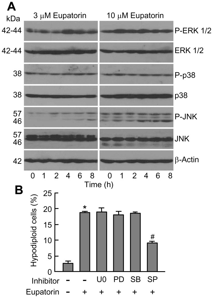
Figure 5. Eupatorin induces phosphorylation of MAPKs.
(A) Time- and concentration-dependent phosphorylation of MAPKs by eupatorin in HL-60 cells. Representative blots are shown. (B) Cells were pretreated with U0126 (U0, 10 µM), PD98059 (PD, 10 µM), SB203580 (SB, 2 µM) and SP600125 (SP, 10 µM) for 1 h and then incubated with eupatorin for 24 h and hypodiploid cells were quantified by flow cytometry. * indicates P<0.05 for comparison with untreated control, # indicates P<0.05 for comparison with eupatorin treatment alone.

