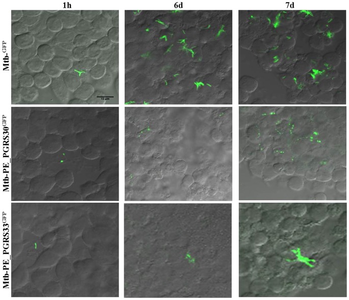
Figure 7. Polar localization of PE_PGRS30 during Mtb macrophage infection.
Macrophages (J774) were infected with the Mtb GFP, Mtb-PEGRS30GFP and Mtb-PE_PGRS33GFP and cells washed and fixed at 1 h and 6 days post-infection. Supernatants from infected macrophages at 6 days post-infection were harvested and used to infect fresh J774 macrophages, that 1 day later were washed and harvested. Slides containing infected macrophages harvested at the different time-points were analyzed at the confocal microscopy and images were obtained using a 63× objective.

