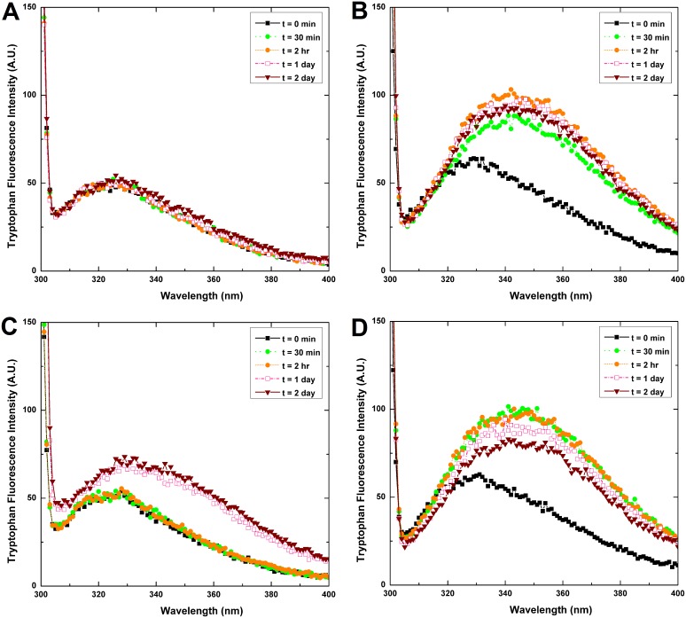
Figure 5. Tryptophan fluorescence intensity measurement of HγD-crys samples (0.1 mg/mL) under different incubation conditions.
(A) HγD-crys incubated at pH 7.0, 37°C; (B) HγD-crys incubated at pH 2.0, 37°C; (C) HγD-crys incubated at pH 7.0, 55°C; and (D) HγD-crys incubated at pH 2.0, 55°C. The fluorescence spectra between 300 and 400 nm were recorded upon exciting the samples at 295 nm. Incubation under pH 2.0 settings resulted in a rapid and greater increase in fluorescence emission, as well as a more noticeable red-shift in the wavelengths of emission maximum than those of the samples under pH 7.0.

