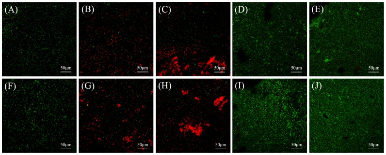
Figure 4. Representative CLSM images of Live/Dead-stained biofilms on material surfaces.
Representative CLSM images of Live/Dead-stained biofilms after 24 h of anaerobic growth on the tested material surfaces: (A) negative control material, (B) positive control material, (C) experimental material, (D) MTA, and (E) Dycal. Biofilms on the corresponding aged samples are shown in (F)–(J). Live bacteria exhibited green fluorescence, and bacteria with compromised membranes exhibited red fluorescence. Scale bars, 50 µm.

