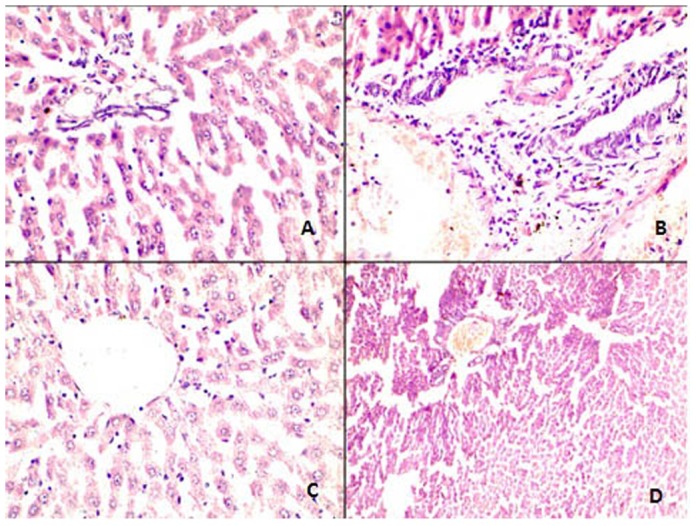
Figure 6. Comparative changes noted on histology- Normal Vs Abnormal at H&E 400 X.

(A) Normal portal triad comprising of hepatic duct, artery and portal vein without any inflammatory infiltrate.; (B) Portal tracts showing mild to moderate periportal inflammation by mononuclear inflammatory cells; (C) Normal central vein which is surrounded by unremarkable looking hepatocytes maintaining their normal trabecular architecture and orientation; (D) Central vein is dilated and congested and shows loss of normal trabecular architecture along with focal necrosis.
