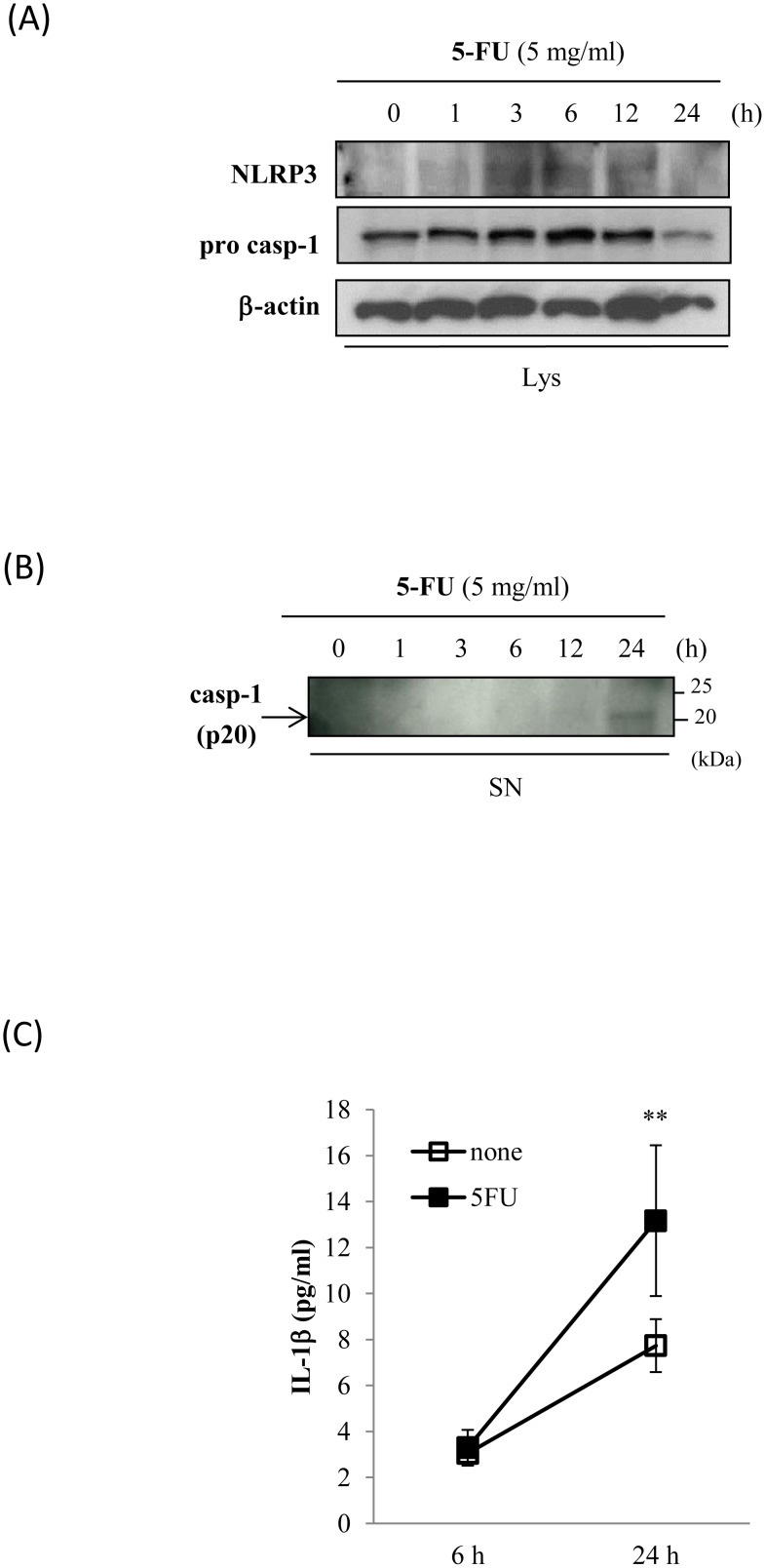
Figure 2. 5-FU-activated inflammasome pathway.
Sa3 cells were incubated with 5 mg/mL of 5-FU for 0 to 24 h. (A), Western blot analysis of the expression of NLRP3 and the precursor of caspase-1 (pro-casp-1) in cell lysates. (B), Western blot analysis of cleaved caspase-1 (p20) in supernatants. Arrow indicates p20-specific bands. (C), ELISA assay of IL-1β in supernatants of Sa3 cells incubated without (open box) or with (closed box) 5 mg/mL of 5-FU for 6 h and 24 h. Values are means ± S.E.M. (n = 4). **p<0.01 compared to Sa3 cells incubated without 5-FU.

