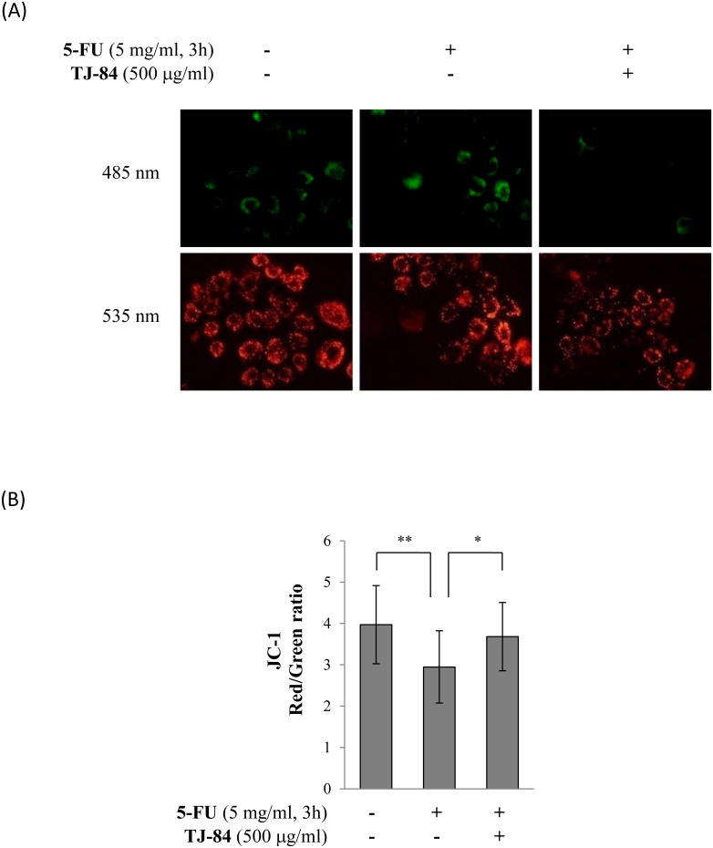
Figure 5. TJ-84 attenuated 5-FU-induced mitochondrial depolarization.
(A), Sa3 cells were incubated with or without 5 mg/mL of 5-FU for 3 h following a 1-h pre-incubation with 500 µg/mL of TJ-84. JC-1 (1 µg/mL) was then loaded for 30 min. JC-1 aggregates (red) and monomers (green) were detected by fluorescence microscopy. (B), The fluorescence intensity per cell was calculated using ImageJ. The calculation of the red/green ratio is shown on the graph. Values are means ± S.E.M. (n = 20, 16, 14). **p<0.01, *p<0.05 compared to the control cells.

