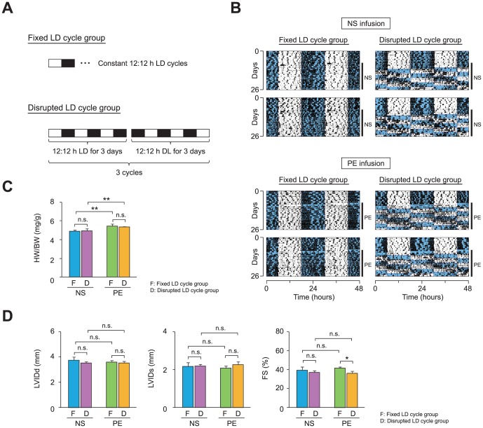
Figure 5. Circadian desynchronization reduces cardiac function in C57BL/6J mice with drug-induced cardiomyopathy.
(A) A light-dark (LD) cycle regimen used to examine the effects of a variable LD schedule on cardiac function. C57BL/6J mice were either maintained on a constant LD schedule (fixed LD cycle group) or were subjected to a 12-h phase shift in LD cycle every 3 days (disrupted LD cycle group). To compare the effects of the LD schedule between animals with healthy hearts and those with cardiomyopathy, either normal saline (NS) or phenylephrine (PE) was continuously infused via an osmotic pump in each LD cycle group. (B) Locomotor activity records of NS-infused (top panel) and PE-infused (bottom panel) mice. Two representative records from animals subjected to the fixed (left column) and disrupted (right column) LD cycle are shown. Activity counts are indicated by the vertical black marks. The records are double-plotted such that 48 h are shown for each horizontal trace. The blue shaded and unshaded areas indicate the dark and light period, respectively. The duration of NS or PE infusion is indicated by the vertical line at the right margin. (C) The ratios of heart weight to body weight (HW/BW) were increased by the PE infusion but were not influenced by the disruption in LD cycle (n = 5–8 per group). (D) The effects of PE, disrupted LD cycle, or both on ventricular function were evaluated using echocardiographic measurements (n = 5–8 per group). LVIDd, LVIDs, and FS are shown in bar graph format. Data are the mean ± SEM. *P<0.05, **P<0.01, two-way ANOVA.

