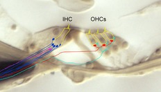
Figure 1. Cross-section of the organ of Corti (3-week-old rat cochlea – unstained).

Hair cells are outlined in yellow, stereocilia and innervation schematized. Type I (blue) and type II (turquoise) afferents contact inner and outer hair cells, respectively. Type II afferents travel hundreds of micrometres toward the cochlea base before contacting hair cells (not shown). Medial (red) and lateral (fuchsia) efferents contact outer hair cells and the dendrites of type I afferents, respectively. Only a single lateral efferent is shown for clarity; in reality a rich plexus of endings is formed beneath each inner hair cell.
