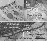
Figure 3. Efferent synapses on mouse outer hair cells.

A, cochlear cross-section showing OHCs from three rows (OHC1–3). B, higher power view of multiple efferent terminals on one wild-type OHC. C, high magnification showing parallel membranes that demarcate the synaptic cistern in apposition to an efferent terminal (reproduced from Fuchs et al. 2014, with permission).
