
Figure 4. Demarcation and reconstruction of an efferent synapse on a mouse OHC.
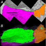
A, single cross-section with the efferent terminal in magenta, the synaptic cistern in green and afferent boutons in umber. B, Z-axis projection (tilted forward ∼30 deg from the plane of section) from a serial reconstruction of 29 sections including that in A (same scale). Same colour scheme as in A with hair cell membrane shown in grey lines. A presynaptic ribbon (turquoise) and associated vesicles (yellow) face the afferent bouton (reproduced from Fuchs et al. 2014, with permission).