
FIGURE 4.
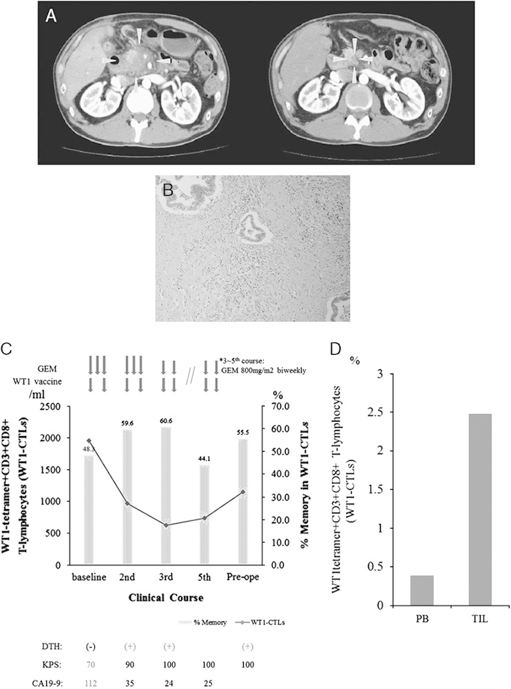
Clinical course and immunologic monitoring of 1 patient. A, Abdominal computed tomography (CT) scan before and after treatment. Left: CT scan at baseline showed a large hypodense lesion in the head of the pancreas, which had also invaded the supramesenteric artery and portal vein. Right: 5 months after treatment (before operation), a follow-up CT scan showed >80% regression of the primary lesion. Gray arrows shows primary lesion of pancreas. B, Microscopic findings of the resected specimen (hematoxylin-eosin stain). C, Clinical course and immunologic monitoring. The black line represents the absolute number of WT1 tetramer+CD3+CD8+ T lymphocytes (WT1-CTLs), and the gray column represents the percentage of memory-phenotype WT1-CTLs. D, Percentages of WT1-CTLs in the peripheral blood (PB) and tumor-infiltrating lymphocytes (TIL). CA19-9 indicates carbohydrate antigen 19-9; CTLs, cytotoxic T lymphocytes; DTH, delayed-type hypersensitivity; GEM, gemcitabine; KPS, Karnofsky performance status.