Fig. 2.
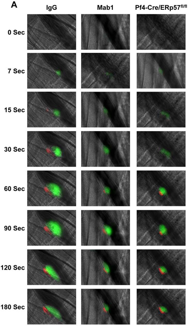
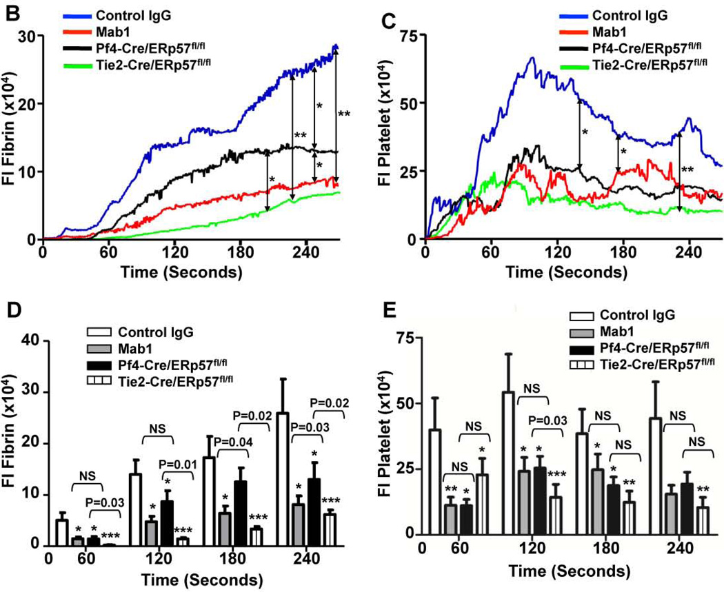
Intravascular ERp57 is required for fibrin deposition and platelet accumulation in vivo. Cremaster arteriole injury was induced in Cre-negative ERp57fl/fl mice pretreated with control IgG or Mab1 (450 µg/mouse) and Pf4-Cre/ERp57fl/fl mice. Platelets and fibrin were detected using anti-CD41 F(ab)2 fragments (binds to platelet β3) conjugated to Alexa Fluor 488 and anti-fibrin antibody conjugated to Alexa Fluor 647. (A) Using intravital microscopy representative fluorescence images show platelet accumulation (green) and fibrin deposition (red) at selected time points up to 180 seconds after vascular injury. Platelet accumulation was visualized at 7 seconds, earlier then the appearance of fibrin, which was seen at 15 seconds. The median integrated fluorescence intensities (FI) of anti-fibrin (fibrin, B) and anti-CD41 (platelet, C) antibodies over 270 seconds were obtained from a total of 30 thrombi in 3 mice per group. Fluorescence signal was not observed using fluorescently labeled control IgG (not shown). The data was analyzed by area under curve (AUC) with a Mann-Whitney rank sum [19]. Only significant differences (*P < .05; **P < .01) are shown. The data was also analyzed by comparing the fluorescence intensity with control mice of anti-fibrin (D) and anti-CD41 (E) antibodies at 60, 120, 180, 240 seconds after vascular injury as described [37]. *P < .05; **P < .01, ***P < .001. P values between the Mab1 and Pf4-Cre/ERp57/fl/fl mice are as indicated.


