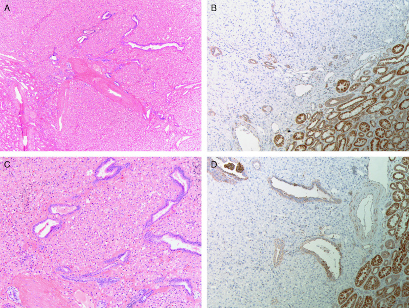
FIGURE 4.

Serial sections stained with hematoxylin and eosin (A and C) and SDHB IHC (B and D). Frequently entrapped benign tubules were noted at the edge of the tumors. SDHB IHC demonstrates positive staining in the internal controls (including the entrapped benign tubules) but all the neoplastic cells are negative.
