
Figure 2.
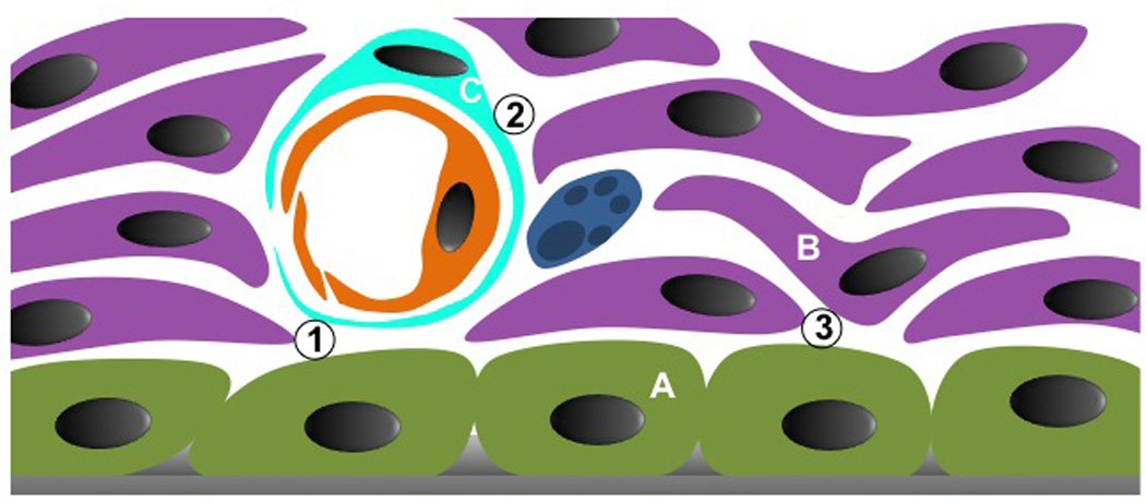
Schematic magnification of the endosteal and subendosteal regions, which compose the endosteal niche. At least, three different stromal cell types can be identified in the niche: A, osteoblasts; B, non-perivascular reticular cells, probably mostly pre-osteoblasts; C, perivascular reticular cells, probably mesenchymal stem cells. These cells will organize the HSC niche. Three interfaces must be considered: 1, osteoblast and perivascular cells; 2, perivascular cells and pre-osteoblasts; and 3, osteoblasts and pre-osteoblasts. Each stromal combination will create a distinct niche for the same HSC pool. How this is organized and what is the dynamic among them is still unknown.