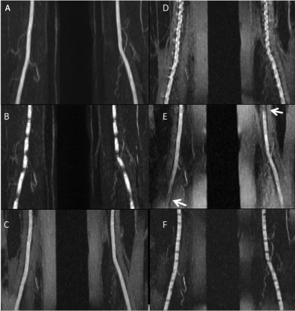
Figure 2.
40 mm-thick maximum intensity projections for QISS and UnQISS acquired in a healthy subject with RR ~760 ms. QISS was acquired with the peripheral vascular coil and fat saturation, sampling bandwidth = 646 Hz/pixel and 92 views. UnQISS was acquired using the body coil without fat suppression, sampling bandwidth 651 Hz/pixel and 316 views. (a) ECG-gated QISS shows uniform arterial signal. (b) QISS (QI = 228 ms) acquired without ECG gating shows horizontal banding artifacts. (c) UnQISS acquired with shot TR/QI = 2400 ms/1200 ms, radial k-space trajectory and a view angle increment (9.68353 degrees, m = 17) shows the arteries comparably to ECG-gated QISS (although with higher fat signal since phase-based fat suppression was not used). (d) UnQISS acquired with identical imaging parameters to (c) except for using a Cartesian k-space trajectory shows severe, artifactual vessel irregularities. (e) UnQISS acquired with identical imaging parameters to (c) except for using a golden view angle increment (111.24612 degrees) shows loss of arterial signal (arrows) in the lower left and upper right corners of the image. (f) UnQISS acquired using shorter shot TR/QI values of 1716 ms/25 ms (TI = 625 ms) shows horizontal banding artifacts.

