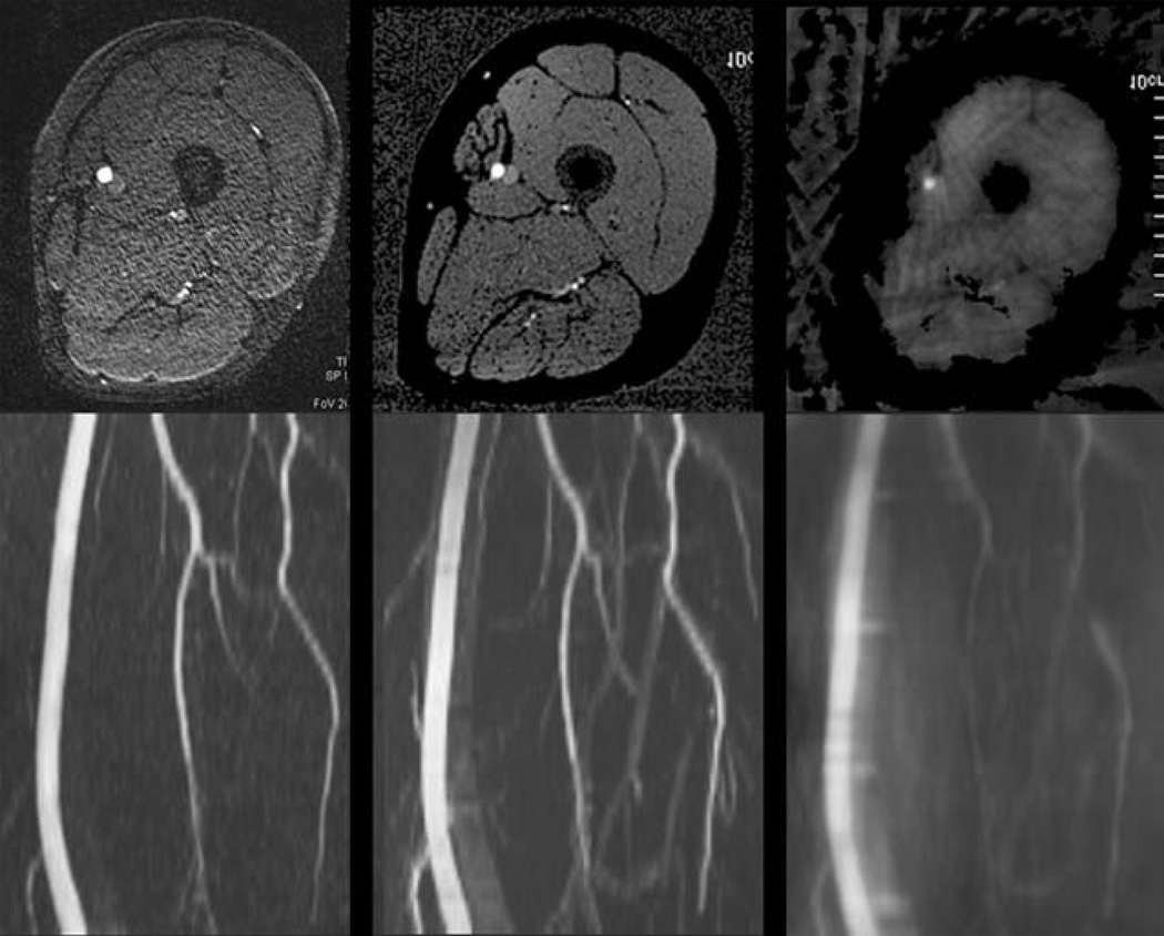
Figure 3.
Impact of azimuthal view angle increment. Top row: Axial images; bottom row: corresponding MIP images. (Left) ECG-gated Cartesian QISS image with 92 views; (middle) UnQISS image with 316 views using an optimal azimuthal view angle increment (9.68353 degrees, m = 7); (right) UnQISS image using a non-optimal view angle increment (9.47368 degrees) shows severe artifacts from non-uniform radial k-space sampling. All images were obtained using phased array coils; UnQISS used phase-based fat suppression.

