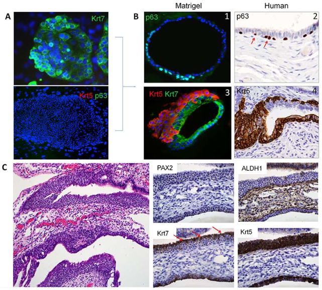
Figure 3.
In vitro and in vivo basal cell differentiation in the oviduct. (A) Colonies of Krt7p/Krt5n/p63n cells from a 20 week old fetal oviduct. (B1, B3) single (p63, green) and multilayered (Krt5, red) basal cell outgrowth seen in matrigel cultures. (B2, B4) similar basal cell growth highlighted by p63 and Krt5 in the adult fimbria. (C) Walthard cell nest in the adult tube is typically PAX2 and ALDH1 negative. Residual Krt7-positive cells (arrows) are displaced from beneath by an expanding Krt5 population.

