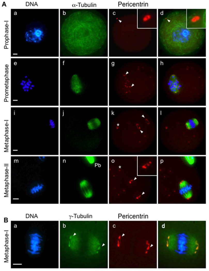
Figure 1.
Pericentrin localizes to MTOCs in oocytes arrested at prophase-I and during meiotic division. (A) Mouse oocytes were collected and fixed for immunofluorescence analysis at specific stages, including prophase-I arrest (a–d, n= 45), prometaphase (e–h, n=51), metaphase-I (i–l, n=65), and metaphase-II (m–p, n=63). The oocytes were double-labeled with specific anti-pericentrin (red, arrows) and anti-acetylated α-tubulin (green) antibodies to detect spindle microtubules. (B) Pericentrin (c, red) co-localizes with γ-tubulin (b, green) specifically at the spindle poles (arrows) in metaphase-I oocytes (n=55). All slides were counterstained with DAPI to detect DNA (blue). Pb: Polar body. *, cytoplasmic MTOCs. Insets show 2× and 4× magnifications of the spindle-pole area. Scale bars, 10 µm.

