Figure 3.
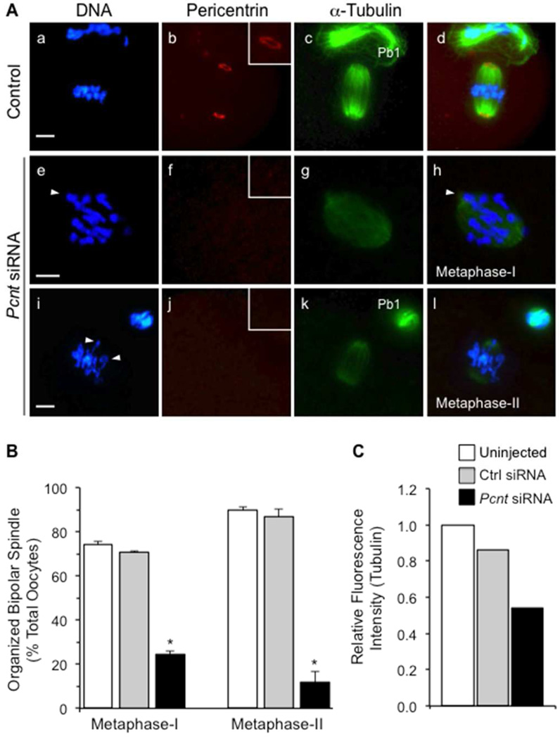
Knockdown of pericentrin leads to the disruption of spindle organization and significant chromosome misalignment. (A) Representative images of the meiotic spindle structure in uninjected (n=137) and non-specific siRNA (a–d, n=110) control oocytes, relative to the Pcnt-siRNA-injected group (e–l, n=113). Oocytes were double-labeled with anti-pericentrin (red) as well as anti-acetylated α-tubulin antibodies for the detection of microtubules (green). DNA is shown in blue and arrows denote misaligned chromosomes. Insets show a 2× magnification of the spindle pole area (arrows). Pb1: First polar body. Scale bar, 10 µm. (B) Percentage (mean ± standard error) of metaphase-I and -II oocytes in each group that exhibit an organized bipolar meiotic spindle structure. * P<0.05. (C) Relative fluorescence (pixel) intensity of acetylated α-tubulin (microtubules) in the spindle of metaphase-I oocytes (n=15 per group). The mean fluorescence intensity values from the uninjected control group was standardized to 1.0, and compared to oocytes injected with non-specific control or Pcnt siRNAs.

