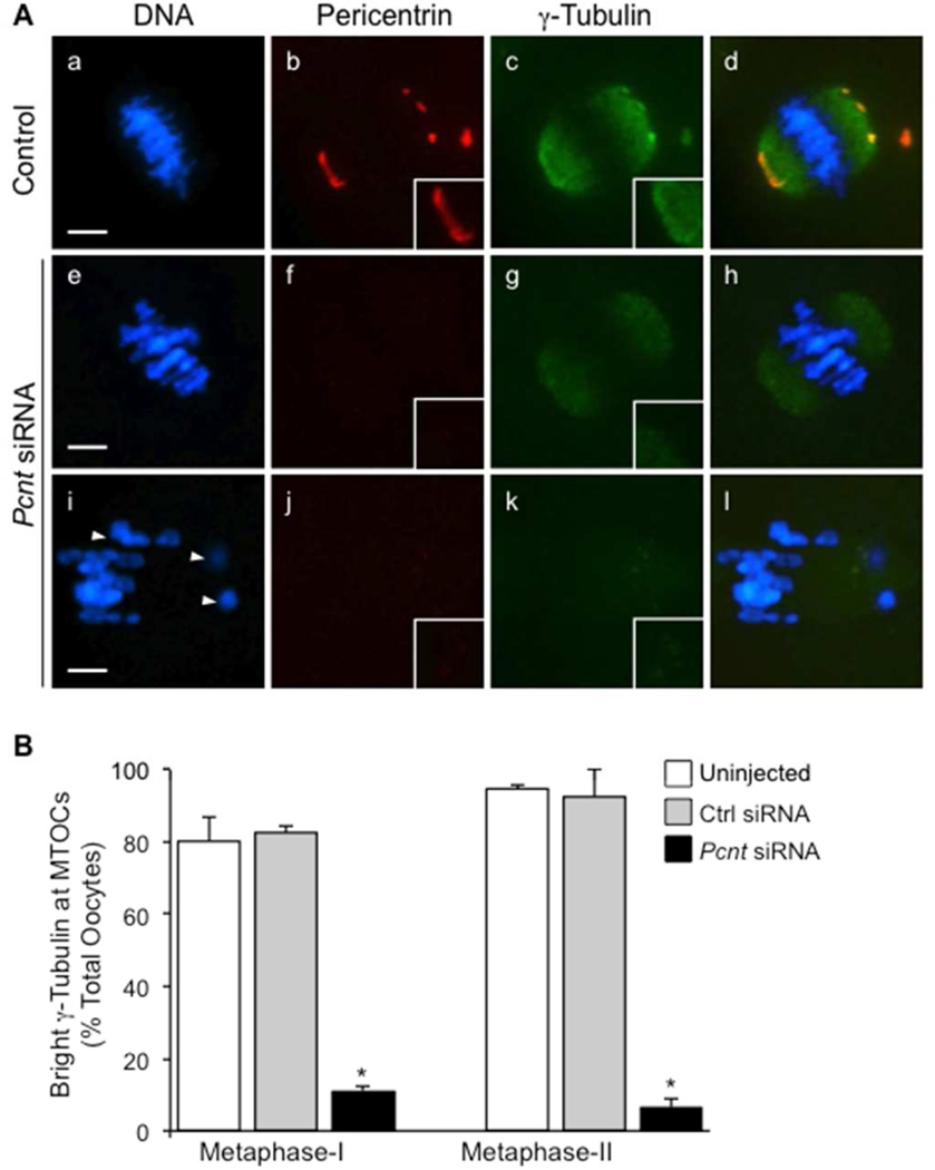
Figure 4.
Knockdown of pericentrin in oocytes decreased γ-tubulin localization at MTOCs. (A) Representative images of oocytes are shown from the uninjected (n=73) and non-specific siRNA (n=89) controls (a–d) compared to the Pcnt-siRNA group (e–h, n=87). Oocytes were double-labeled with anti-pericentrin (red) and γ-tubulin (green) antibodies. Arrows denote misaligned chromosomes. (B) The percentage (mean ± standard error) of oocytes with bright γ-tubulin detected at MTOCs was lower in the Pcnt-siRNA group relative to the uninjected and non-specific siRNA controls. DNA is shown in blue. * P<0.05. Insets show a 2× magnification of the spindle pole area. Scale bars, 10 µm.

