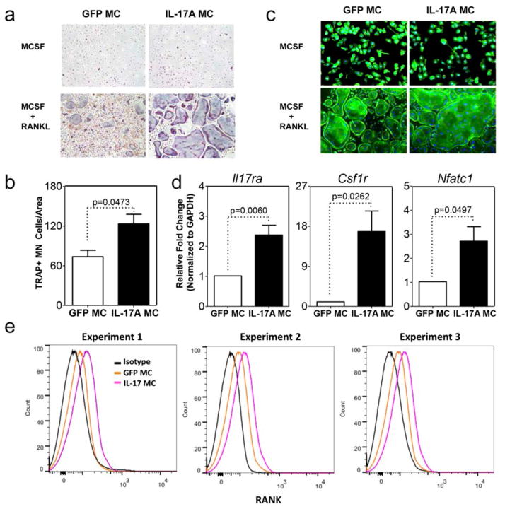
Figure 3. IL-17A induces osteoclast differentiation in a RANKL dependent manner in vitro.
TRAP a) cytochemical stain and b) quantitative analysis of TRAP+ multinucleated (MN) cells CD11b+ sorted bone marrow macrophages cells from GFP or IL-17A MC injected mice cultured for 4 days with M-CSF and RANKL. c) F-actin ring formation assay. Data for panel a and b are pooled from 4 experiments using 3 mice per group. (d) qPCR of sorted RANK+ cells for expression of II17ra, csf1r and Nfatc1 normalized to Gapdh (data are pooled from 2 experiments using 3 mice per group). e) Flow cytometric analysis of RANK expression on cells extracted from bone marrow after GFP or IL-17A MC injection. Histograms are from 3 independent experiments. Black denotes isotype control, orange denotes GFP MC, and pink denotes IL-17A MC.

