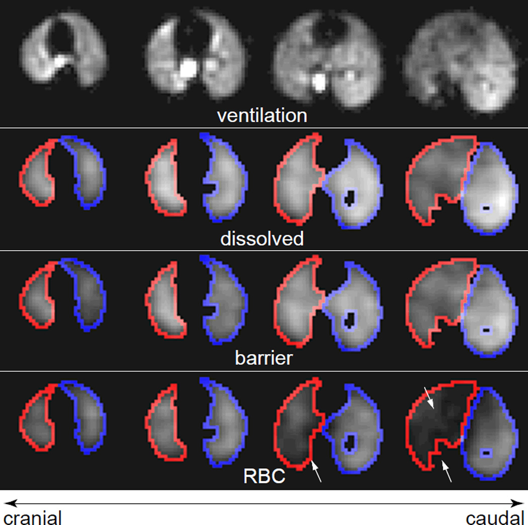
Figure 5.
Axial slices from a 3D HP 129Xe MR images of a Bleomycin-treated rat. (a) Ventilation image. Again, the lung parenchyma is reasonably well ventilated. Also shown are the corresponding slices from the (b) total dissolved 129Xe image, (c) barrier tissue image, and (d) RBC image. Outlines depict the edges of the mask used to confine data analysis to the injured (right, red) and uninjured (left, blue) lung. Arrows indicate regions of reduced RBC signal within the injured lung.

