Figure 1.

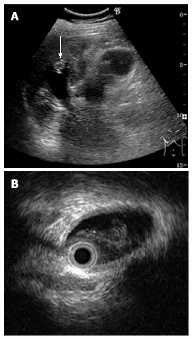
Ultrasonogram demonstrating a hyper-echoic lesion (arrow) in a dilated posterior bile duct of the right lobe (A), Intraductal ultrasonography demonstrates the presence of hypo-echoic material, that was considered likely to be mucin (B).

Ultrasonogram demonstrating a hyper-echoic lesion (arrow) in a dilated posterior bile duct of the right lobe (A), Intraductal ultrasonography demonstrates the presence of hypo-echoic material, that was considered likely to be mucin (B).