Figure 1.
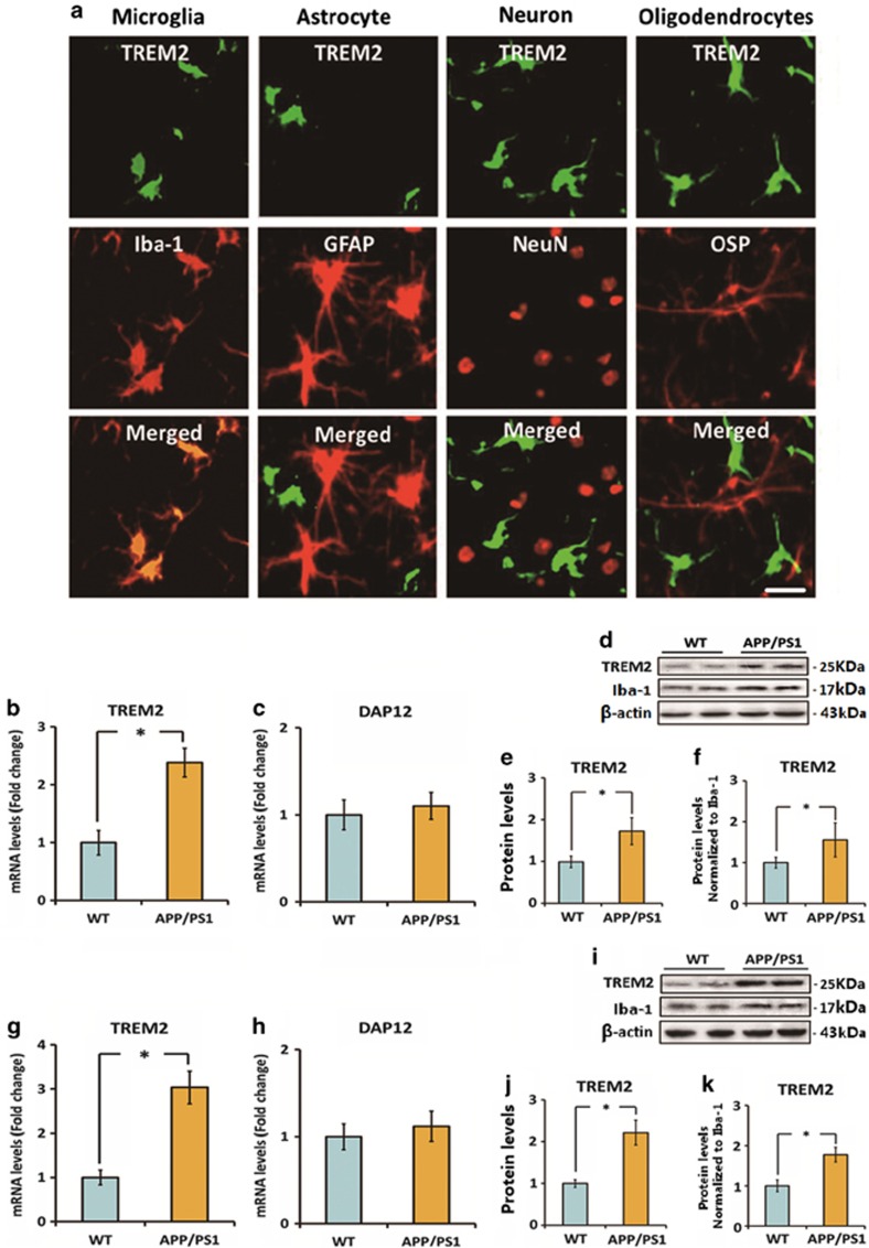
Triggering receptor expressed on myeloid cells 2 (TREM2) is localized on microglia and is upregulated under Alzheimer's disease (AD) context. (a) The cellular localization of TREM2 in the brain of 7-month-old APPswe/PS1dE9 mice was investigated by double immunofluorescence staining. TREM2 was colocalized with Iba-1 (a microglia marker) rather than GFAP (an astrocyte marker), NeuN (a neuron marker), and OSP (an oligodendrocyte marker). Scale bar=20 μm. (b and c) mRNA levels of TREM2 and DAP12 in the cerebral cortex of 7-month-old APPswe/PS1dE9 mice and wild-type (WT) mice were measured by quantitative reverse transcription-PCR (qRT-PCR). Data were normalized to the levels of GAPDH mRNA. (d–f) Protein levels of TREM2 in the cerebral cortex of 7-month-old APPswe/PS1dE9 mice and WT mice were detected by western blot analysis. Data were normalized to β-actin or a microglia marker Iba-1. (g and h) mRNA levels of TREM2 and DAP12 in the hippocampus of 7-month-old APPswe/PS1dE9 mice and WT mice were measured by qRT-PCR. Data were normalized to the levels of glyceraldehyde 3-phosphate dehydrogenase (GAPDH) mRNA. (i–k) Protein levels of TREM2 in the hippocampus of 7-month-old APPswe/PS1dE9 mice and WT mice were detected by western blot analysis. Data were normalized to β-actin or a microglia marker Iba-1. All data were analyzed by independent sample t-test. Columns represent mean±SEM (n=8 per group). *P<0.05. It should be noted that cropped gels are used in this figure, and the full-length gels are shown in Supplementary Figure S6.

